Overview
-

-
Overview
-
Laboratory Introduction
Laboratory Introduction
TUMOR MICROENVIRONMENT LABORATORY
TUMOR MICROENVIRONMENT LABORATORY
Tumor microenvironment is a unique environment in tumors whereby various types of cells including immune cells, endothelial cells, fibroblasts, and adipocytes are interacting each other to survive along with cancer cells. Metabolic alterations including hypoxia and glycolysis (aerobic and anaerobic), and its consequence, which is acidic environment are all part of the tumor microenvironment. We are interested in understanding how tumor microenvironmental components are interacting each other (such as how immune cells are behaving in hypoxic/acidic environment; how macrophages consume oxygen and glucose making tumors more hypoxic and glycolytic) to make tumors more resistant towards anticancer therapies. To do this, we utilize various cutting edge technologies such as fluorescence-activated cell sorting, in vivo techniques with various GEMM (genetically engineered mouse models), and in vitro biochemical analysis techniques.
Related Researcher

G-ONE AHN Professor
- Email : goneahn@snu.ac.kr
Research topics
① It is well known that tumor-associated macrophages (TAM, the most frequent immune cell population in tumors) promote the tumor progression. We have learned that cancer cells consume oxygen and glucose to produce energy. However, TAM are also cells, so shouldn’t TAM also consume oxygen and glucose? If so, how would they co-operate with cancer cells? In our recent research, we found that TAM compete with cancer cells for the oxygen and glucose making tumors more hypoxic and glycolytic. Importantly, with collaboration with clinicians at SNUH, we observed that NSCLC (non-small cell lung cancer) patients with high FDG (fluorine deoxyglucose) PET (positron emission tomography) signals contained higher numbers of TAM and vice versa. We are now expanding this research to other tumor types and other roles that TAM may play in modulating the tumor metabolism. ② We are particularly interested how tumor microenvironment regulates tumor response to radiotherapy (since hypoxia is an ultimate resistance factor to radiotherapy). In our latest paper, we were able to experience the latest radiotherapy technique, called FLASH radiotherapy, developed by Prof. Billy Loo at Stanford University. They’re delivering high dose of ionizing radiation at the ultra-fast speed (at least 20 times faster than conventional radiotherapy technique), lowering the normal tissue toxicity significantly. We found that FLASH irradiation also produces very interesting changes in the tumor microenvironment compared to conventional irradiation. ③ We are also interested in investigating how macrophages sense hypoxia. Cells usually activate a transcription factor called hypoxia-inducible factor (HIF) under hypoxic conditions to turn on many genes involved in survival/death/cell cycle arrest, adaptation to hypoxia, and angiogenesis. In our lab, we have knockout mice targeting HIF pathways only in macrophages. In our recent papers, we have challenged these mice to cerebral ischemia (stroke model through middle cerebral artery ligation), hindlimb ischemia (through femoral artery ligation), and inflammatory bowel disease to study interesting and disease-specific role of HIF in macrophages. We are now investigating the role of HIF in macrophages in regulating diet-induced obesity.
Research goals
To identify new targets in tumor microenvironment improving therapeutic response of tumors. To conduct translational research by understanding deeper level of molecular changes in tumors treated with radiotherapy.
Research achievements
Kim YE, Gwak SH, Hong BJ, Oh JM, Choi HS, Kim MS, Oh D, Lartey F, Rafat M, Schuler, E, Kim HS, von Eyben R, Weissman IL, Koch CJ, Maximi PG, Loo BW, Ahn GO. Effects of ultra-high dose rate FLASH irradiation on the tumor microenvironment in Lewis lung carcinoma: role of myosin light chain. Int J Radiat Oncol Biol Phys. 2020 In Press
Lim HS, Lee Y, Heo J, Heong H, Hong KT, Kwon DH, Shin MH, Oh M, Sable GA, Ahn GO, Lee JS, Song HK. Targeted degradation of transcription coactivator Src-1 through the N-degron pathway. Angew Chem Int Ed Engl. 2020, Sep 28;59(40):17548-17555
Jeong HB, Kim SH, Hong BJ, Lee CJ, Kim YE, Bok SY, Oh JM, Gwak SH, Yoo MY, Lee MS, Chung SJ, Defrene J, Tessier P, Pelletier M, Jeon HR, Roh TY, Kim BJ, Kim KH, Ju JH, Kim SE, Lee YJ, Kim DW, Kim IH, Kim HJ, Park JW, Lee YS, Lee JS, Cheon GJ, I L Weissman, Chung DH, Jeon YK, Ahn GO. Tumor-Associated Macrophages Enhance Tumor Hypoxia and Aerobic Glycolysis. Cancer Res. 2019 Feb 15;79(4):795-806.
Kim YE, Lee MJ, Gu HJ, Kim JW, Jeong SJ, Yeo SJ, Lee YJ, Im SH, Sung YC, Kim HJ, I L Weissman, Ahn GO. HIF-1α activation in myeloid cells accelerates dextran sodium sulfate-induced colitis progression in mice. Dis Model Mech. 2018 Jul 30;11(7):dmm033241.
Bok SY, Kim YE, Woo YS, Kim SE, Kang SJ, Lee YT, Park SK, I L Weissman, Ahn GO. Hypoxia-inducible factor-1α regulates microglial functions affecting neuronal survival in the acute phase of ischemic stroke in mice. Oncotarget. 2017 Dec 1;8(67):111508-111521.
Hong BJ, Kim JW, Jeong HB, Bok SY, Kim YE, Ahn GO. Tumor hypoxia and reoxygenation: the yin and yang for radiotherapy. Radiat Oncol J. 2016 Dec;34(4):239-249.
Kim KS, Jeon SU, Lee CJ, Kim YE, Bok SY, Hong BJ, Park DY, Ahn GO, Kim HJ. Radiation-Induced Esophagitis In Vivo and In Vitro Reveals That Epidermal Growth Factor Is a Potential Candidate for Therapeutic Intervention Strategy. Int J Radiat Oncol Biol Phys . 2016 Jul 1;95(3):1032-1041.
Song CH, Hong BJ, Bok SY, Lee CJ, Kim YE, Jeon SR, Wu HG, Lee YS, Cheon GJ, Paeng JC, D J Carlson, Kim HJ, Ahn GO. Real-time Tumor Oxygenation Changes After Single High-dose Radiation Therapy in Orthotopic and Subcutaneous Lung Cancer in Mice: Clinical Implication for Stereotactic Ablative Radiation Therapy Schedule Optimization. Int J Radiat Oncol Biol Phys . 2016 Jul 1;95(3):1022-1031.
Bok SY, Wang TJ, Lee CJ, Jeon SU, Kim YE, Kim JW, Hong BJ, Yoon CJ, Kim SJ, Lee SH, Kim HJ, Kim IH, Kim KH, Ahn GO. In vivo imaging of activated microglia in a mouse model of focal cerebral ischemia by two-photon microscopy. Biomed Opt Express . 2015 Aug 7;6(9):3303-12.
Ahn GO, Jun Seita, Hong BJ, Kim YE, Bok SY, Lee CJ, Kim KS, Lee J C, N J Leeper, J P Cooke, Kim HJ, Kim IH, I L Weissman, J M Brown. Transcriptional activation of hypoxia-inducible factor-1 (HIF-1) in myeloid cells promotes angiogenesis through VEGF and S100A8. Proc Natl Acad Sci U S A . 2014 Feb 18;111(7):2698-703.
Ahn GO, D Tseng, Liao CH, M J Dorie, Agnieszka Czechowicz, J M Brown. Inhibition of Mac-1 (CD11b/CD18) enhances tumor response to radiation by reducing myeloid cell recruitment. Proc Natl Acad Sci U S A . 2010 May 4;107(18):8363-8.
Ahn GO, J M Brown. Matrix metalloproteinase-9 is required for tumor vasculogenesis but not for angiogenesis: role of bone marrow-derived myelomonocytic cells. Cancer Cell . 2008 Mar;13(3):193-205.
Ahn GO, K J Botting, A V Patterson, D C Ware, Moana Tercel, W R Wilson. Radiolytic and cellular reduction of a novel hypoxia-activated cobalt(III) prodrug of a chloromethylbenzindoline DNA minor groove alkylator. Biochem Pharmacol . 2006 Jun 14;71(12):1683-94.
Ahn GO, D C Ware, W A Denny, W R Wilson. Optimization of the auxiliary ligand shell of Cobalt(III)(8-hydroxyquinoline) complexes as model hypoxia-selective radiation-activated prodrugs. Radiat Res . 2004 Sep;162(3):315-25.
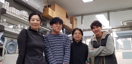
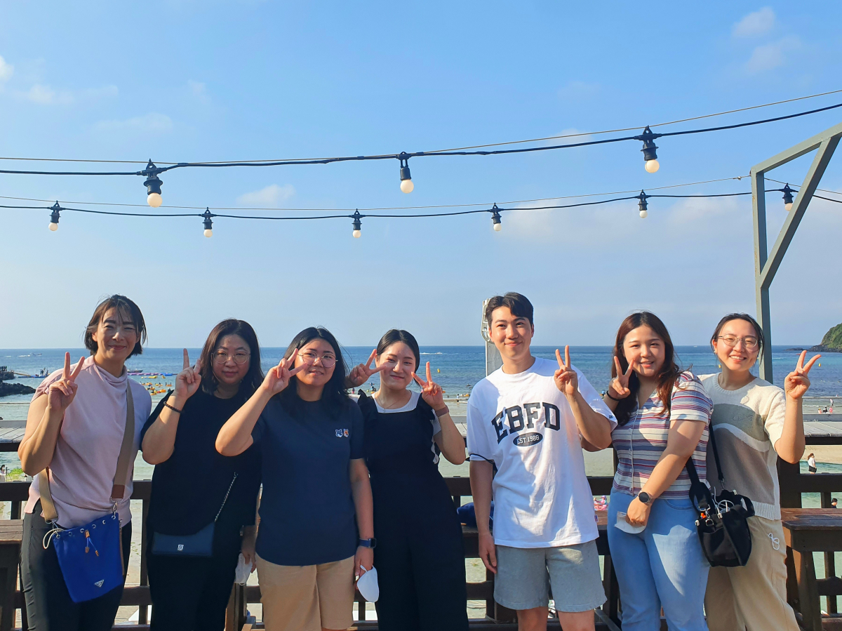
Photos