Overview
-

-
Overview
-
Laboratory Introduction
Laboratory Introduction
Artificial Intelligence Digital Twin Lab (AIDTL)
Artificial Intelligence Digital Twin Lab (AIDTL)
In medical field, the importance of the technologies for medical imaging analysis and application based on artificial intelligence (AI) have become enlarged as the medical benefits and usefulness of these technologies have been approved. Particularly, AI enables us not only to develop and assess biomarkers of lesions based on big data in medical images but also to classify severity as well as to predict prognostic factors; it is necessary that this research has to be performed by research labs professionally being in charge of imaging engineering. Since this research lab was launched in September 2011, the lab has given its priority to the AI technology, which takes lead to advanced medical technologies, and the research diversely utilizing high technologies, such as 3D printing, augmented reality (AR), and metaverse, has been actively ongoing for the opening the new future of medicine in conquest of COVID19 and cancers. The research lab for AI imaging technology and 3D printing has been collaborating with many researchers from Seoul National University Hospital for the development of the future medical IT technologies and leading to the change of medical paradigm as continuously attempting innovative medical trials. This research lab is consisting of AI research division, SW Research & Development division, 3D printing division, and AR technology division, and the lab maximizes its research efficiency via close relations in research development as wells as the close communication between researchers and clinicians.
Related Researcher

Jin Mo Goo Professor
- Email : jmgoo@snu.ac.kr
Research topics
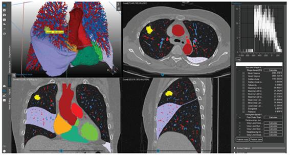
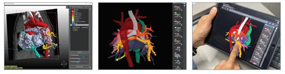
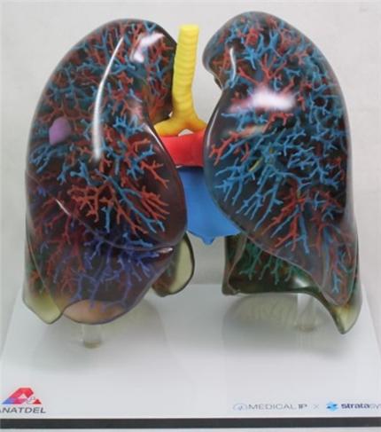
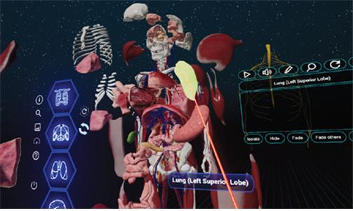
Main research topics / research accomplishment 1) The development of technology for AI-based medical imaging analysis: On the basis of the segmentation technologies of medical images based on machine learning and deep learning, anatomical data segmentation and analysis technologies for reading medical images and surgical support are developed. The key technology is that, using AI, each of the body parts, such as organs, legions, and body compositions, is segmented fast and accurately. 2) The development of human organ models for surgical simulation based on patient-specific imaging data: The research collaborating with many researchers within the hospitals are ongoing by using 3D printed organ models approved by MFDS as medical devices. The medical images that are accurately detected and segmented using AI technology can be linked with the web-based mobile platforms through the mesh construction providing more intuitive 3D models. The accessibility on mobile platforms makes researchers communicating in real time. 3) The development of medical contents of metaverse related to VR/AR technology: The AR/VR anatomical contents using the data of actual patients are developed, and these are linked with 3D virtual world on metaverse. Thus, it is possible to do hands-on medical education with advanced technology, and the cadavers used for the anatomic education in medical schools can be substituted.
Research goals
The research lab for AI imaging technology and 3D printing strives continue the advancement in high medical IT technologies and discover unmet needs and solve the problems by communicating with researchers from diverse fields of medicine. Moreover, the lab will become a leader of the development in global medical industry through clinical adaptation of various medical IT technologies.
Research achievements
Automated Lung Segmentation on Chest Computed Tomography Images with Extensive Lung Parenchymal Abnormalities Using a Deep Neural Network. Korean J Radiol. 2020;21:e163. doi: 10.3348/kjr.2020.0318.2020-10-01 PMID: 33169549
Differentiation of left atrial appendage thrombus from circulatory stasis using cardiac CT radiomics in patients with valvular heart disease. Eur Radiol, Cardiac, Published: 19 August 20202020-08-19 PMID : 32812175
Anterior Pulmonary Ventilation Abnormalities in COVID-19. Radiology. 2020 Nov;297(2):E276-E277. doi: 10.1148/radiol.2020203043. Epub 2020 Aug 13.2020-08-13 PMID: 32787702
Prognostic Implication of Volumetric Quantitative CT Analysis in Patients with COVID-19: A Multicenter Study in Daegu, Korea. Korean J Radiol. 2020;21:e130. English. Published online Aug 04, 2020.2020-08-04 PMID: 32767868
Extension of Coronavirus Disease 2019 (COVID-19) on Chest CT and Implications for Chest Radiograph Interpretation. Radiology: Cardiothoracic Imaging. Vol. 2, No. 2 (Mar 30 2020)2020-03-30
Application of computerized 3D-CT texture analysis of pancreas for the assessment of patients with diabetes. PLoS One. 2020 Jan 13;15(1):e0227492. doi: 10.1371/journal.pone.0227492. eCollection 2020.2020-01-13P MID: 31929591
Advanced Medical Use of Three-Dimensional Imaging in Congenital Heart Disease: Augmented Reality, Mixed Reality, Virtual Reality, and Three-Dimensional Printing. Korean J Radiol. 2020;21:e6. English. Published online Jan 08, 2020.2020-01-08 PMID: 31997589
Relationships between Spinal Sarcopenia and Spinal Sagittal Balance in Older Women. Annals of Geriatric Medicine and Research . 2019 Sep;23(3):141-148. doi: 10.4235/agmr.19.0030. Epub 2019 Sep 25.2019-09-25 PMID : 32743302
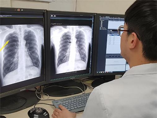
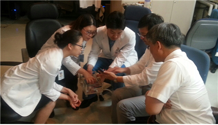
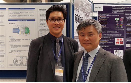
Photos